

What is Diagnostic Musculoskeletal Ultrasound?
Diagnostic musculoskeletal ultrasound is an advanced imaging technology that uses high-frequency sound waves to produce real-time images of muscles, tendons, ligaments, nerves, joints, bursae, and other soft tissues
Unlike X-rays, ultrasound does not use radiation making it safe for EVERYONE. Unlike MRI, ultrasound allows structures to be examined while you move, making it one of the most valuable imaging tools for evaluating many musculoskeletal injuries.
Diagnostic Musculoskeletal Ultrasound
See Beneath the Surface. Understand the Problem More Clearly.
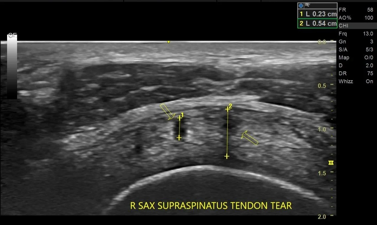
This diagnostic ultrasound image from a one of Dr.Dinh’s patients indicate tears of the supraspinatus tendon in the shoulder , measuring approximately 0.23 cm and 0.54 cm.
High-resolution musculoskeletal ultrasound allows Dr. Bryan Dinh to visualize tendon integrity in real time, helping identify the precise location and extent of injury while correlating findings with your symptoms and physical examination.
DID YOU KNOW?
Unlike MRI, diagnostic ultrasound allows Dr. Dinh to compare the injured side with the healthy side immediately during your examination. This real-time comparison improves diagnostic confidence and helps distinguish normal anatomical variation from true pathology
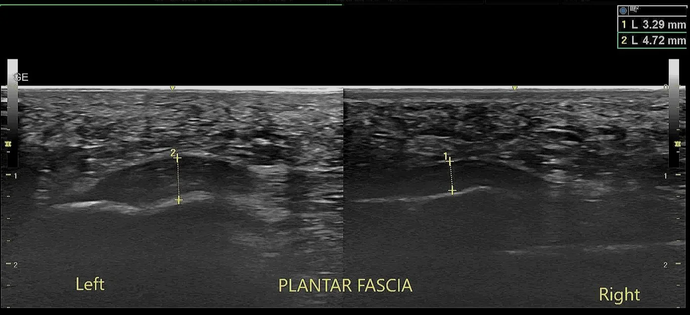
This ultrasound indicate a thicker plantar fascia in the symptomatic left foot (4.72 mm) compared with the healthy right foot (3.29 mm), consistent with plantar fasciopathy.
Comparing both sides is one of the unique advantages of diagnostic ultrasound, allowing Dr. Bryan Dinh to identify subtle tissue abnormalities and tailor treatment based on objective findings.
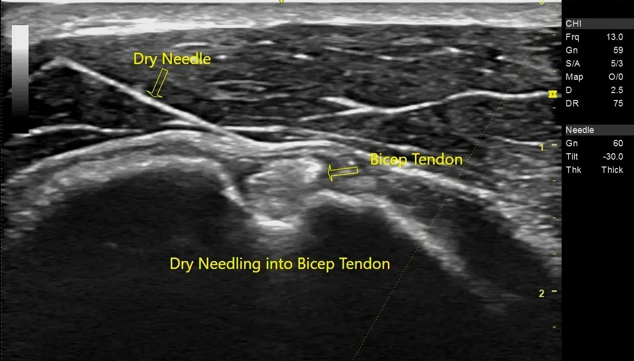
The image shown demonstrates Dr.Dinh performs real time ultrasound-guided regenerative dry needling of a pathological long-head biceps tendon. Real-time imaging confirms accurate needle placement throughout the procedure, improving precision while protecting adjacent tendons, nerves, and blood vessels.
From Precision Diagnosis to Precision Intervention
“Diagnostic ultrasound is not the end of the evaluation—it becomes the foundation for treatment.”-Dr.Dinh
Traditional physical therapy often relies on physical examination and anatomical landmarks to estimate where treatment should be applied.
At Iron Tiger Physical Therapy, we take a different approach.
Once the source of pain has been identified, high-resolution musculoskeletal ultrasound allows treatment to be delivered precisely to the injured tissue.
This integration of advanced imaging with targeted intervention represents a level of musculoskeletal care rarely available within physical therapy practices.
Why Iron Tiger?
High-resolution diagnostic ultrasound is only as valuable as the clinician interpreting it.
Dr. Bryan Dinh is among the very small number of physical therapists in Oklahoma who hold the Registered in Musculoskeletal Sonography (RMSK®) credential—a globally recognized certification demonstrating advanced expertise in musculoskeletal ultrasound around the world.
Combined with fellowship training in orthopedic manual physical therapy (FAAOMPT), this allows your evaluation and treatment to be guided by both advanced imaging and expert clinical reasoning, creating a level of precision that is rarely available in outpatient physical therapy.
FAQ
-
Not always. Ultrasound excels at evaluating muscles, tendons, ligaments, bursae, and peripheral nerves in real time. MRI remains the preferred study for some conditions, such as deep intra-articular pathology, bone marrow abnormalities, or certain complex injuries.
-
Diagnostic ultrasound requires extensive additional education beyond physical therapy school.
Interpreting ultrasound images accurately requires advanced knowledge of anatomy, pathology, physics, image optimization, and clinical reasoning.
The Registered in Musculoskeletal Sonography (RMSK®) credential is considered one of the highest certifications in musculoskeletal ultrasound and requires rigorous examination.
Dr.Dinh is only one of the two PT that has RMSK® credential in Oklahoma
-
Integrity, creativity, and empathy shape the way we work. These aren't just words—they’re the foundation of everything we build.
-
Yes.
One of the greatest advantages of diagnostic ultrasound is immediate interpretation.
Dr. Dinh reviews the images with you during your appointment, explains what is normal and abnormal, and discusses how the findings relate to your symptoms and treatment plan.
-
Can ultrasound show inflammation?
Sometimes.
Ultrasound can identify several indirect signs associated with inflammation, including:
Fluid accumulation
Synovitis
Bursal swelling
Increased tendon thickness
Altered tendon structure
-
Most imaging studies provide only a single static snapshot.
Ultrasound allows your tissues to be examined while moving.
This means Dr. Dinh can evaluate structures during:
Shoulder elevation
Squatting
Walking
Running
Throwing
Lifting
Neck rotation
Tendon gliding
Nerve movement
-
Diagnostic Ultrasound excels at:
Tendon injuries
Tendinopathy
Muscle tears
Ligament injuries
Peripheral nerve entrapments
Dynamic instability
Bursitis
Joint effusions
Real-time movement assessment
Ultrasound-guided procedures
MRI excels at:
Bone marrow abnormalities
Deep cartilage injuries
Meniscal pathology
Labral tears
Stress fractures
Certain spinal conditions
Tumors
Complex intra-articular pathology
For many common orthopedic conditions, diagnostic ultrasound has demonstrated diagnostic accuracy comparable to MRI while providing the additional benefit of dynamic examination.
-
Yes.
High-resolution musculoskeletal ultrasound has excellent diagnostic accuracy for many tendon injuries, particularly:
Rotator cuff tears
Achilles tendon tears
Quadriceps tendon injuries
Patellar tendon injuries
Distal biceps tendon tears
Gluteal tendon tears
Ultrasound can often determine:
Whether a tear is partial or complete
Tear location
Tear size
Tendon retraction
Tissue quality
-
Yes.
Modern musculoskeletal ultrasound is increasingly used to evaluate peripheral nerves.
Examples include:
Carpal tunnel syndrome
Cubital tunnel syndrome
Radial tunnel syndrome
Tarsal tunnel syndrome
Common peroneal neuropathy
Meralgia paresthetica
Ultrasound can evaluate:
Nerve enlargement
Compression
Dynamic subluxation
Scar tissue
Relationship to surrounding anatomy
This information complements electrodiagnostic studies when indicated